
MRI Cavum Septum Pellucidum et Vergae Stock Image C043/0388 Science Photo Library
DOI: 10.7759/cureus.57907 Corpus ID: 269059080; Symptomatic Cavum Vergae Cyst in a Geriatric Patient: A Report of a Rare Case and Conservative Management Approach @article{Penchev2024SymptomaticCV, title={Symptomatic Cavum Vergae Cyst in a Geriatric Patient: A Report of a Rare Case and Conservative Management Approach}, author={Plamen Penchev and Petar-Preslav Petrov and Vladislav Velchev and.

Abnormalities Associated With the Cavum Septi Pellucidi on Fetal MRI What Radiologists Need to
During development, these spaces obliterate anteroposteriorly: the cavum vergae followed by the cavum septum pellucidum. Some researchers believe that the development of cavum septum pellucidum and the development of cavum vergae are linked, but this is still debatable [2, 20].If the cavum septum pellucidum and cavum vergae are present, an endoscopic transforaminal approach into the third.

Cavum septi pellucidi and cavum vergae MedLink Neurology
Background Cavum septum pellucidum (CSP) and cavum vergae (CV) are normal anatomical variations present in some children, adolescents, and adults. When the cavity is larger than normal, it is.

Cavum septum pellucidum, cavum vergae, and cavum veli interpositi (annotated CT) Image
If there is an extension posterior to a vertical plane created by the columns of the fornix, the term cavum vergae is used. 6,39 The clinical significance of cavum septi pellucidi and cavum vergae is unknown, and they are considered a normal variant. 39 However, studies have suggested that their presence is associated with cognitive dysfunction.

Cavum vergae Ars Neurochirurgica
Cave of septum pellucidum seen on CT. The cave of septum pellucidum (CSP), cavum septi pellucidi, or cavity of septum pellucidum is a slit-like space in the septum pellucidum that is present in fetuses but usually fuses during infancy. The septum pellucidum is a thin, laminated translucent vertical membrane in the midline of the brain separating the anterior horns of the right and left ventricles.

Cavum Septi Pellucidi and Cavum Vergae With Increased Amyloid β Cortical Load in a 65YearOld
The septum pellucidum (meaning translucent wall in Latin - SP), also known as the ventricle of Sylvius, is a thin, triangular double membrane separating the frontal horns of the right and left lateral ventricles of the brain. It extends between the anterior portion of the corpus callosum, and the body of the fornix and its width varies from 1.5 to 3.0 mm

Absent Cavum Septi Pellucidi American Journal of Obstetrics & Gynecology
The anatomist Sylvius of Leyden University first described cavum septi pellucidi in 1671 (07). Cavum Vergae is a posterior extension of the cavum septi pellucidi, communicating with the cavum septi pellucidi but lying posterior to the columns of the fornix (70). In the early literature, these cavities were considered to be the fifth and sixth.

Cavum septi pellucidi Ars Neurochirurgica
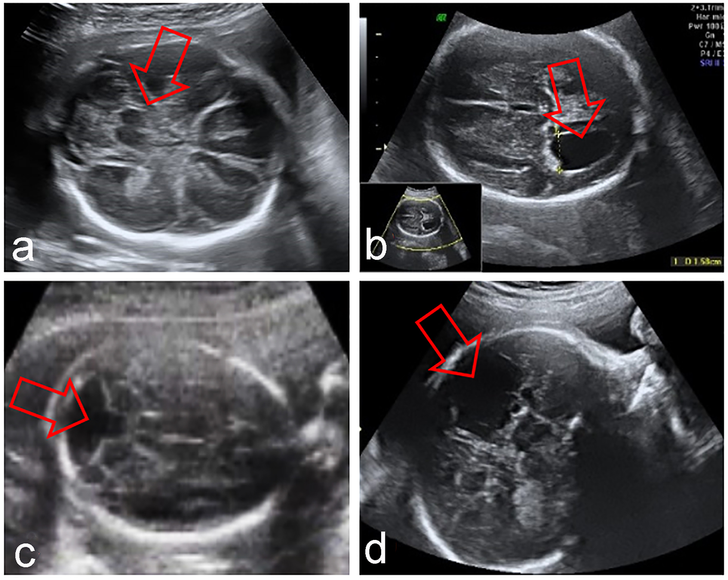
Cavum septi pellucidi and cavum vergae in normal and developmentally delayed populations.. 12. Ho YK, Turley M, Marc-Aurele KL, Jones MC, Housman E, Engelkemier D, et al.. Enlarged cavum septi pellucidi and vergae in the fetus: a cause for concern. J Ultrasound Med 2017; 36: 1657-68. doi:.

Cavum Septi Pellucidi (CSP) Brain imaging
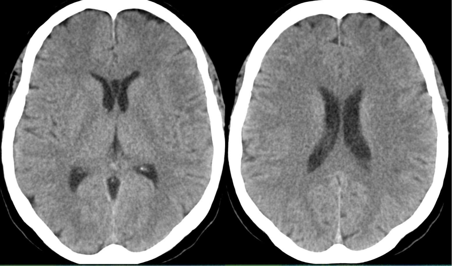
Age: 45 years. Gender: Female. ct. Evidence of a rectangular shaped CSF containing space between the leaflets of the septum pellucidum (between the anterior horns of lateral ventricles) with posterior extension splaying the fornices laterally representing an incidental cavum septum pellucidum with cavum vergae (cavum septum pellucidum et vergae).

Abnormalities Associated With the Cavum Septi Pellucidi on Fetal MRI What Radiologists Need to
During development, these spaces obliterate posteroanteriorly - the cavum vergae followed by the cavum septum pellucidum - and it is not uncommon that both occur together as one contiguous space (see cases 3 and 4), aptly termed "cavum septum pellucidum (or septi pellucidi) et vergae". Boundaries. anterior: genu of the corpus callosum
of Fetuses With Abnormal Cavum Septi Pellucidi Experience of a Tertiary Center Zorila
Cavum septum pellucidum (CSP) is a common incidental finding, defined as a midline cerebrospinal fluid (CSF) space delimited superiorly by the crus of the fornices and inferiorly by the tela choroidea of the third ventricle [].It is anatomically distinct from cavum vergae (CV) which is a CSF space extending posteriorly to the columns of the fornix.

Abnormalities Associated With the Cavum Septi Pellucidi on Fetal MRI What Radiologists Need to
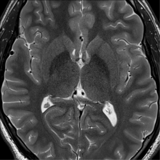
Purpose: The morphology of the cavum septi pellucidi (CSP), cavum Vergae (CV), and cavum veli interpositi (CVI) has been infrequently explored with neuroimaging. The aim of the present study was to delineate these cavities using magnetic resonance (MR) imaging. Methods: A total of 71 patients were enrolled in the present study. . Following initial examinations with conventional MR sequences.

Cavum Septi Pellucidi (CSP) Brain imaging
Purpose The morphology of the cavum septi pellucidi (CSP), cavum Vergae (CV), and cavum veli interpositi (CVI) has been infrequently explored with neuroimaging. The aim of the present study was to delineate these cavities using magnetic resonance (MR) imaging. Methods A total of 71 patients were enrolled in the present study. Following initial examinations with conventional MR sequences.
Cava septi pellucidi et vergae Image
The width of the cavum septi pellucidi was greater than the width of the cavum vergae at every gestational stage with both structures attaining their maximum width around 29-31 gw (Figs. 8 and and9). 9). After that time, there was a reduction in width, most prominently in the cavum vergae, although a cavity was present in all foetuses in the.
Cavum septum pellucidum, cavum vergae, and cavum veli interpositi (annotated CT) Image
Cavum septum pellucidum (CSP) cyst, cavum vergae, and cavum velum interpositum are various presentations of benign midline anterior intracranial cysts. They are pathological when symptomatic, which arise depending upon the size of the cysts. Cavum septum pellucidum cysts are rare lesions with an incidence of 0.04% . Symptomatic cysts of CSP are.

🥇 Anatomía del SEPTUM PELLUCIDUM. ¡Explicación Rápida y Sencilla! YouTube
Cavum septum pellucidum et cavum vergae Cavum veli interpositi; 3 articles feature images from this case. Cavum septum pellucidum; Cavum vergae; Cavum veli interpositi; 29 public playlists include this case. GK - Neuro - Anatomy by GLK; Head and neck by Robert Ligetfalvi;
- Wie Viele Geschwister Hat Jesus
- سورة ق مكتوبة كاملة بالتشكيل
- How Much Time I Wasted On Lol
- Led Spiegel Mit Uhr Und Bluetooth
- Birnen Bohnen Und Speck Tim Mälzer
- Stadt In Italien 5 Buchstaben Beginnend Mit A
- Fahren Die Busse In Wuppertal
- المسلسل التركي حكاية جزيرة مدبلج
- For Your Eyes Only Abkürzung
- Vhs Osnabrück Land Programm 2024